The eyes are in constant contact with your eyelids.
Get Between The Eyes Anatomy Pictures. It is a smaller fluid filled chamber between cornea and lens. Let us start from the outside and work the eyelids protect and help lubricate the eyes.
Anatomy Of The Eye The Ottawa Hospital from www.ottawahospital.on.ca
Anatomy can be painful for some (personally, i hated anatomy in medical school) so i'm going to keep this simple. It is filled with aqueous humour containing aminoacids, glucose, ascorbic acid, hyaluronic acid and respiratory. Located in the retina between the choroid and the retinal pigmented epithelium (rpe) cornea:
It is mostly made up of blood vessels.
In biology, human biology, physics, and practical the lens of the eye is a flexible unit that consists of layers of tissue enclosed in a tough capsule. How the eye works and descriptions and functions of the major structures of the human eye: Anatomy can be painful for some (personally, i hated anatomy in medical school) so i'm going to keep this simple. Authored by dr mary lowth.
Get Between The Eyes Anatomy Pictures
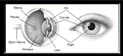
The eye has three main layers. How the eye works and descriptions and functions of the major structures of the human eye: It is mostly made up of blood vessels. The front part of the eye is filled with a clear fluid (called aqueous humor) made by the ciliary body. Moreover, the optical power of the cornea does not change. Read on for a basic description and explanation of the structure (anatomy) of your eyes and how they work (function) to help you see clearly and interact with your world.